Ultrasound
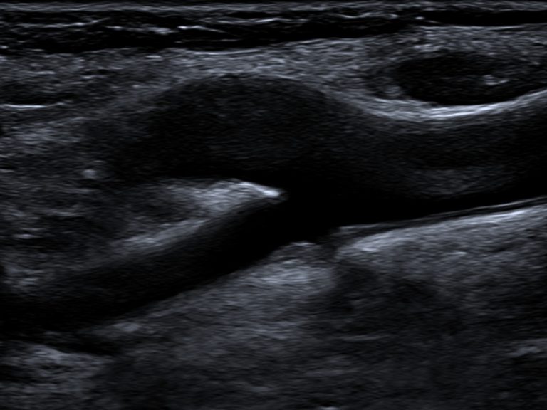
An ultrasound scan is a procedure that uses high frequency sound waves to generate images of internal body structures. Ultrasound does not use ionising radiation so is considered very safe.
To acquire the images water-based gel is applied to your skin and then a hand-held ultrasound transducer is moved over the area of interest.
The ultrasound examination is performed by our highly trained Sonographers who then forward the images to our specialist doctors. The Radiologist then prepares a report for your referring practitioner.
Diagnostic Ultrasound examinations often have specific preparation requirements. Please see below for general information about preparing for Ultrasound examinations. Your personal preparation requirements will be outlined at the time of booking.
Abdomen / Abdomen Doppler / Renal arteries
Do not eat, smoke or chew gum for 6 hours before appointment. Regular sips of water are recommended to stay hydrated.
Pelvis / Renal / 1st Trimester pregnancy
90 minutes prior to the appointment, empty your bladder and then drink one litre of water, finishing water 1 hour prior to exam. A FULL BLADDER is required.
2nd Trimester pregnancy
90 minutes prior to the appointment, empty your bladder and then drink one litre of water, finishing water 1 hour prior to exam. You may empty up to 30 minutes prior to exam.
3rd Trimester pregnancy
Drink 500mls of water 1 hour prior to scan. Empty bladder as required.
Your Sonographer will collect you from the reception area and escort you to the ultrasound room. Depending on your scan you may be required to change into a gown.
The Sonographer will use a transducer to obtain different views and record a series of images. This process is generally pain-free, however some pressure may need to be applied to improve the image. Your Sonographer may request that you also assist in certain scans, for example – holding your breath, or by turning or standing.
Your scan time will vary depending on the complexity of the requested examination. Most simple ultrasound scans take approximately 15-20 minutes, however more time is required for complex studies such as certain obstetric or vascular imaging.
When the scan is complete, it may be necessary for you to wait while our Radiologist reviews the obtained images. Occasionally further imaging may be required. Your Sonographer will advise you if this is the case.
The cost of the scan depends on a number of factors, including the type of scan that your doctor has requested and whether you have a Government issued concession card. Please advise our friendly Customer Service Team if you are a pension or health care card holder.
Our Customer Service Team will be able to advise you of all costs involved with your ultrasound including any out of pocket cost (if relevant).
Many Ultrasound examinations have specific Federal Government Pre-requisites in place to enable you to claim a medicare rebate for that examination. When you book your appointment you will be advised if a rebate is applicable for your examination.
Some categories of examinations commonly asked about are explained on the next few tabs such as Pregnancy and Musculoskeletal (MSK)
When performed on the same day, Medicare will NOT rebate the second examination:
Ultrasound Doppler WITH Ultrasound Musculoskeletal or General or Pregnancy
Ultrasound Pregnancy WITH Ultrasound Pelvis
Ultrasound Abdomen WITH Ultrasound Renal
Ultrasound Pelvis WITH Ultrasound Renal
Ultrasound Renal WITH Ultrasound Abdomen
Ultrasound Musculoskeletal Bilateral
Pregnancy Scans
Pregnancy Scans are subject to Medicare Eligibility Criteria to qualify for a Medicare rebate: For Comprehensive explanation of the criteria and timing of the scans, please visit here
Please note, there is often preparation required for a pregnancy scan and our friendly clerical staff will advise you of this upon booking.
If you wish to obtain a digital copy of your images, please ask our staff to provide you with the steps to activate your digital access.
Pregnancy Scans
To assess fetal risk the examination MUST be performed between 11.5 to 13.5 weeks of pregnancy. Due to this narrow time frame it is essential that patients make an appointment as soon as they receive the referral from their medical practitioner. The associated blood test may be completed any time after 10 weeks and preferably 3 days before the ultrasound.
The RANZCOG guidelines for prenatal assessment can be found here.
Musculoskeletal Ultrasound
US SHOULDER / UPPER ARM
Rebatable Criteria:
• Evaluation of injury to tendon, muscle or muscle/tendon junction; or
• Rotator cuff tear/calcification/tendinosis (biceps, subscapular, supraspinatus, infraspinatus); or
• Biceps subluxation; or
• Capsulitis and bursitis; or
• Evaluation of mass including ganglion; or
• Occult fracture; or
• Acromioclavicular joint pathology
Benefits are NOT payable when referred for non-specific PAIN alone.
US KNEE
Rebatable Criteria:
• Abnormality of tendons or
• bursae about the knee; or
• Meniscal cyst, popliteal fossa cyst, mass or pseudomass; or
• Nerve entrapment, nerve or nerve sheath tumour; or
• Injury of collateral ligaments
Benefits are NOT payable when referred for non-specific knee PAIN alone or other knee condition including:
• Meniscal and cruciate ligament tears
• Assessment of chondral surfaces


