Dental Imaging & OPG
Dental Imaging involves not only looking at the teeth but also the upper and lower jaws, and often the entire face. Your dentist/oral surgeon may also require information on bone growth when planning surgical procedures for younger people.
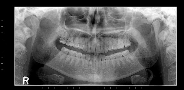
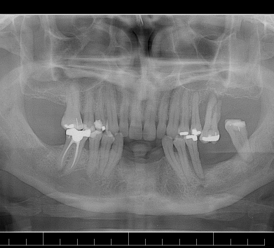
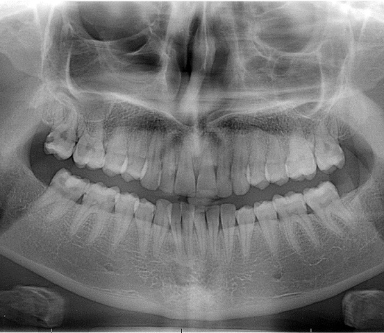
The basic tools of dental imaging are the OPG (Orthopantomogram) and Cephalometry. These x-rays require specialised x-ray machines. An OPG (“orthopantomogram”) gives a panoramic view of the mouth, giving information on the teeth and the bones of the upper and lower jaw. Cephalometry is used to obtain measurements and determine relationships of the structures of the lower face.
The temporo-mandibular joints (TMJ’s) are often the cause of facial symptoms. These are the joints of the lower jaw just in front of the ears. They allow your mouth to open and close. These are imaged using both a standard x-ray technique, and also specialised OPG views.
If an OPG is not possible for whatever reason, or for a detailed assessment of the bones and soft tissues (skin, muscle and glands) of the face, your dentist/oral surgeon may ask for a CT. This test generates cross sections of the face, and with the aid of computer, can produce three-dimensional images of the face.
There is a specialised form of CT called a “Data Scan”, which is used to plan some dental procedures like titanium implant surgery. This uses a standard set of CT images to generate multiple sections through either jaw in different planes. Using these Data Scan images, the bone where the implant is planned can be evaluated for thickness, depth and bone density.
All jewelry from around the head and neck including hair bands. This is because hair pulled together shows up on x-ray impacting the quality of the x-ray; it is not the material the hair bands are made from which degrades the images.
